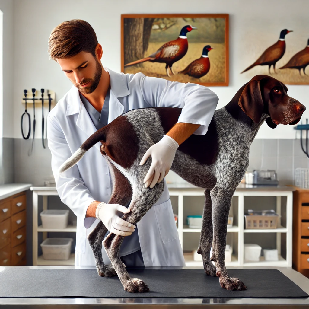
In my initial plan, I intended to save the knee for the final part of this series because, quite frankly, it’s one of the most controversial joints we’ll be discussing. However, as I gave it more thought, it made sense to cover the entire rear end of the dog at this point in the series. So, here we are diving into the complex anatomy of the knee, or stifle joint, and the common injuries associated with it.
Understanding the Stifle Joint
The knee in dogs is a fascinating, complex condylar synovial joint. Sometimes it’s referred to as a hinge joint, but that really only scratches the surface. Structurally, it’s quite similar to our own knees, with the joint formed by the condyles at the end of the femur (thigh bone) connecting with the flat top of the tibia (shin bone).
There are multiple key components that stabilize and function within the knee joint:
- Cruciate Ligaments: These are two ligaments that cross each other (hence the term “cruciate”). In humans, they’re known as the anterior and posterior cruciates, while in dogs, they’re called cranial and caudal. Both essentially serve the same function. The one we commonly associate with injuries is the front (cranial) ligament, often called the ACL in dogs, the term coming from humans.
- Collateral Ligaments: These include the medial and lateral collateral ligaments which help stabilize the knee.
- Patellar Tendon and Patella: The main tendon of the quadriceps, known as the patellar tendon, contains an ossified structure — the patella, or knee cap.
- Menisci: Inside the joint, there are two fibrocartilaginous structures, known as the medial and lateral meniscus, which act as shock absorbers and help even out the articulation.
- Synovial Fluid: The joint space is filled with a thick, lubricating fluid called synovial fluid, which allows the joint to move smoothly.
All these elements work together to allow the knee to function normally. While we often think of the knee as simply hinging in flexion and extension, these structures also allow for some degree of side-to-side movement as well as torsion.
Common Knee Issues in Dogs
While there are numerous potential knee problems, for the sake of brevity, I’ll focus on the two most common issues: luxating patella and cruciate ligament disease (the latter of which we’ll cover in future posts).
1. Luxating Patella
A luxating patella occurs when the knee cap (patella) pops out of its natural groove. Most of us can relax the muscles in our leg and gently move our knee cap around, but even with that movement, it stays within its groove. In dogs with this condition, however, the knee cap dislocates, typically medially (toward the other knee).
What does this look like? You’ll often see a dog suddenly go lame, sometimes hiking their leg up high. In some cases, this acute lameness resolves just as suddenly when the patella slides back into place.
There are two primary ways this condition occurs:
- Developmental: In many cases, the dog’s anatomy is formed incorrectly, such as a groove that’s too shallow or ligament attachments that don’t hold things in place properly. Congenital luxating patella typically causes the knee cap to slip medially.
- Traumatic: Less commonly, a traumatic event can cause the patella to luxate.
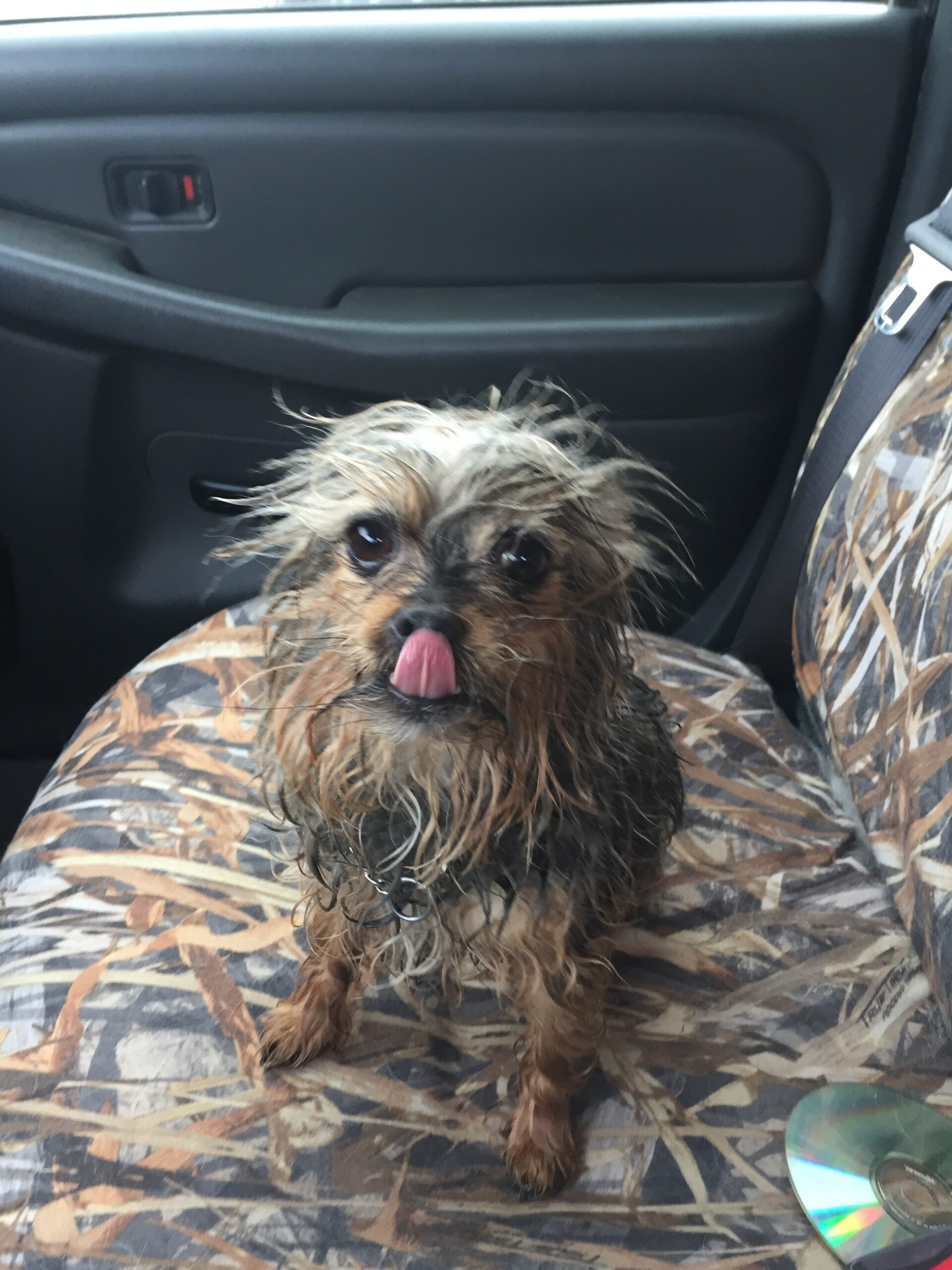
Who’s at risk? This condition is most common in small breeds like Yorkies, Poodles, and Shih Tzus. It also shows up with some regularity in Spaniel breeds. In the larger sporting breeds like Labs and Goldens, it’s less common but does occur.
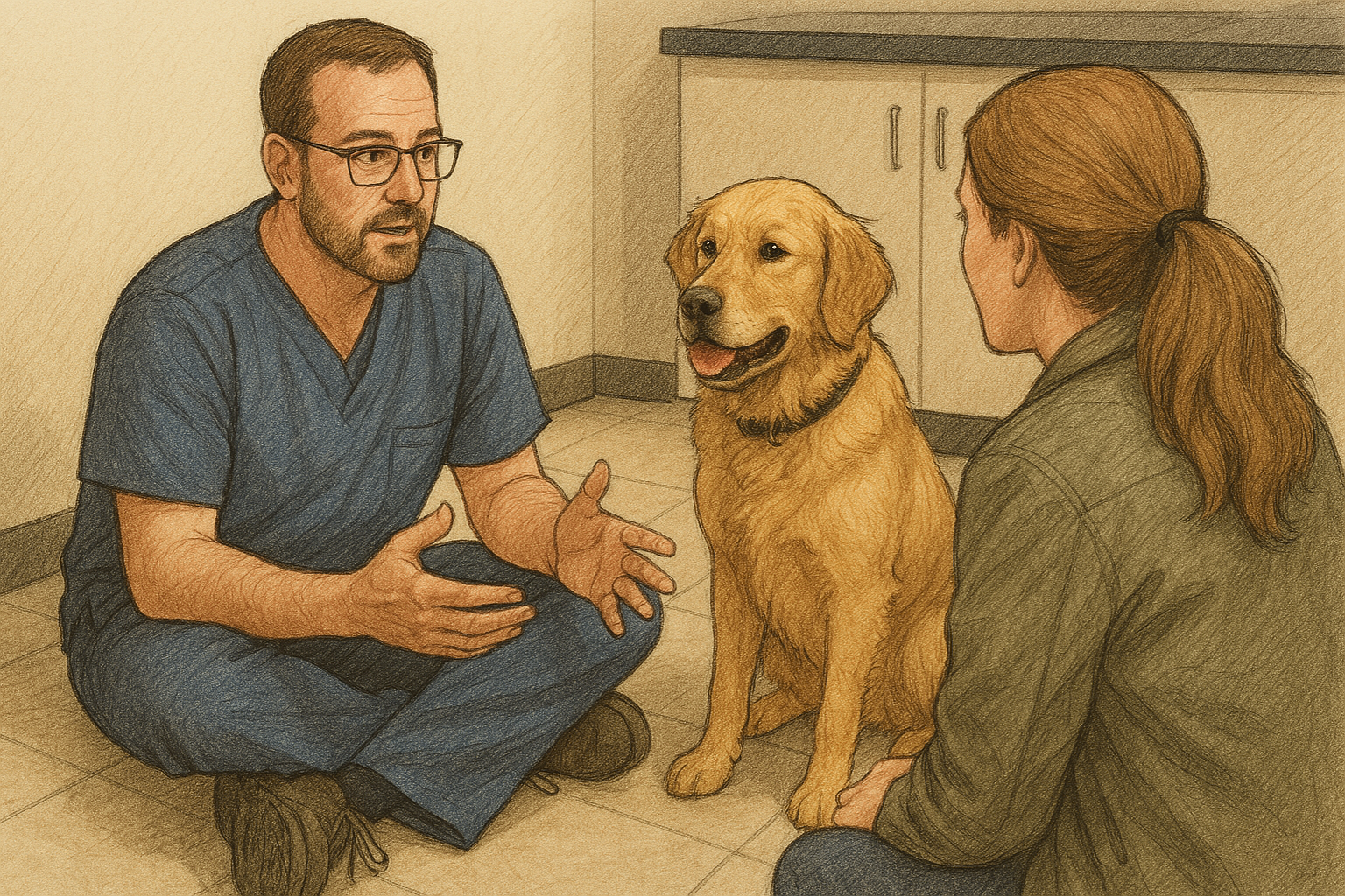
Treatment Options: If a dog shows clinical signs (lameness), surgery is usually the best option, especially if the issue is congenital. In rare instances, particularly with trauma-induced luxation, a rehabilitation program can sometimes help avoid surgery. However, I always set expectations low since targeting the affected limb during rehab can sometimes worsen the condition. If that happens, surgery becomes necessary.
Surgical Correction: This typically involves a combination of procedures like deepening the groove, placing sutures to prevent medial tracking, or even a tibial transposition to adjust the patellar tendon attachment. Most dogs do very well post-surgery, but I’ll add a word of caution: larger dogs are at a higher risk of complications if post-op care isn’t strictly followed.
What’s Next?
I had initially planned to dive into cruciate ligament disease in this post, but as I started writing, I realized that topic deserves its own deep dive (or two). So, stay tuned for the next part where we’ll cover everything you need to know about cruciate ligament issues in dogs.


